Combined Transcatheter Mitral and Tricuspid Valve Repair with Edge to Edge Technique
The Patient
• Elderly female (88), NYHA III dyspnea, active with daily activities
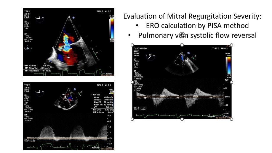
• Severe mitral regurgitation (ERO 43mm 2 , R VOL 60ml)
• Annular dilatation, degenerative leaflet changes, some hypermobility
• Severe tricuspid regurgitation (ERO 80mm 2
•Functional with annular dilatation
•LVEF 60%, non dilated
•Coronaries, 60% mid LAD, conservative Rx
•Pacemaker (6/2019), atrial fibrillation, arthritis, hypertension
•STS for mortality 5.76%
•HEART Team decision for transcatheter intervention, both mitral and tricuspid
TEE: Commissural Mitral View with Color
TEE: Simultaneous Biplane View of Mitral Valve
Commissural and Long Axis
Significant mitral regurgitation due to annular dilatation and hypermobility of both leaflets with some degenerative changes ERO 43mm 2 R VOL 60ml

Severe Functional Tricuspid Regurgitation

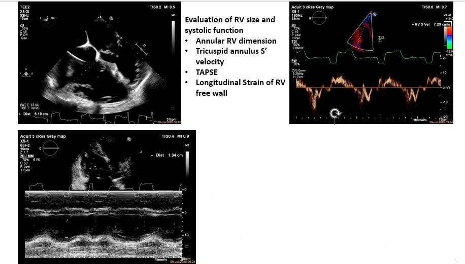
Evaluation of RV size and systolic function
- Annular RV dimension
- Tricuspid annulus S’ velocity
- TAPSE
- Longitudinal Strain of RV free wall
TEE: Transgastric 3D of the Tricuspid Valve
Tricuspid Valve Assessment: Mid-Esophageal Views
Advancement of PASCAL device in left atrium via transeptal puncture and change to closed configuration
Mitral Valve Repair with Pascal (10mm) in A2-P2 area
Note independent clasping of posterior leaflet for optimal result Mild residual MR
Mitral Valve Repair with PASCAL P10 Comparison baseline and final result
Iatrogenic septal defect: Intermittent right to left shunting due to elevated right atrial pressures
Positioning of Pascal for Tricuspid Septal-Anterior Leaflet Clasping
Transgastric Short Axis Tricuspid View: Evaluation of correct orientation of Pascal device
Evaluation of leaflet insertion after closing the Pascal device
Tricuspid Valve Repair with Pascal (10mm) Mild residual TR
Final color flow evaluation of interatrial septal defect: Left to right shunting only, after TV repair
Discharge Images
Color flow evaluation of the mitral valve in apical 4-chamber view
Color flow evaluation of the tricuspid valve in apical 4-chamber view
